Posted on TBNewsWatch September 30, 2023
BY JULIO HELENO GOMES
A Thunder Bay-based company and researchers at Lakehead University are refining technology they developed to spot early-stage breast cancer that may be used to scan other parts of the body. The device now being customized to provide diagnostic images of other organs is a significant advance in cancer screening, says the principal investigator.
“Basically it’s the same underlying technology but it’s the next step in the technology we use for detection of breast cancer,” says Dr. Alla Reznik. “It’s next-generation technology. It’s not just re-configuring because it represents the next evolutionary step.”
Reznik is a professor and the Tier 1 Canada Research Chair in the Physics of Radiation Medical Imaging. Since joining Lakehead in 2008 she has continued her interest in radiation medical imaging technologies and, through the spin-off company Radialis Inc., has created a device to diagnose cancer in women with dense breast tissue where X-ray mammography is not sufficient. With a grant of $740,000 from the Canadian Institute for Health Research, Reznik and her team at Lakehead are upgrading the technology to seek different cancers, including prostate.
“At this stage our technology is customized for imaging in multiple organs, not only the breast,” Reznik says.
“This is an ongoing project, what we call ‘translational’ research, to turn an initial invention of a novel medical imaging technology to innovation. In order to achieve impactful research outcomes we need to navigate the whole journey, of research and development, then find clinical partners to prove the effectiveness of the technology, and then commercialization.”
The goal of this research is to come up with a versatile tool that can target different organs for imaging at high sensitivity and spatial resolution, explains graduate student Anirudh Shahi. Now in the second year of his master’s program, Shahi was attracted to the endeavour because it allows him to apply his physics background to tackle real-world problems in health care. He also appreciates the opportunity to expand his skill-set, working in disciplines ranging from fundamental physics to imaging software. He’s specifically involved in fusing data acquired from different angles around objects to improve the quality of the images generated.
“Such a tool can enable both accurate diagnosis and treatment monitoring of cancer patients in our community in the future,” says Shahi, who was born in India and grew up in Thunder Bay. “On a day-to-day basis, students from the community also benefit since they are given the opportunity to take part in a collaborative learning environment, lead research projects, and collaborate with industry.”
For Brandon Baldassi, there are unique challenges associated with applied physics in medicine.
“The idea of optimizing a complex physical system to address challenges in the diagnosis and treatment of cancer was very compelling,” he says of what drew him to this field.
Baldassi, a Thunder Bay native, studied medical physics at McMaster University. When he participated in Reznik’s lab as a summer student in 2016, he decided to pursue his master’s degree at Lakehead. After graduating this spring he joined Radialis as systems R & D lead and has found this project into organ-targeted positron emission tomography (PET) to be both more challenging and more rewarding than he initially expected.
“This research has the potential for significant clinical impact,” Baldassi states. “The commercialization of this technology offers great opportunity for student interns, new graduates, and local industry to flourish within the medical technology space.”
Reznik’s career in radiation medical imaging research centres on personalized or precision medicine – delivering the right treatment to the right patient at the right time. Personalized treatment needs to be supported with personalized diagnosis, she says. For imaging the breast, her technology addresses the crucial need for personalized diagnostic for women with radiologically dense breast tissue for whom conventional x-ray mammography is ineffective.
“Our organ-targeted technology offers much higher sensitivity so we can reduce the dose of radio pharmaceuticals,” Reznik says, adding that scan time goes from a typical 40 minutes to as little as five minutes. “So the device is more efficient, scanning time is shorter. What does this mean for hospitals? You can scan more patients, which means everything becomes much more cost-efficient.”
The Positron Emission Mammography (PEM) detector designed for breast cancer is a plug-in device the size of a shopping cart, meant to be compact enough to take to under-serviced communities. That technology has already undergone a series of clinical trials, and a second round was expected to take place in September.
Reznik says the market price for their device is $500,000, compared to $3-million for a whole-body PET scanner.
“So everything becomes much more efficient and we can see better, more accurate, more precise images – using lower amounts of radio pharmaceuticals and a lower-cost device,” she says.
Research Matters highlights the important work of researchers at Lakehead University.
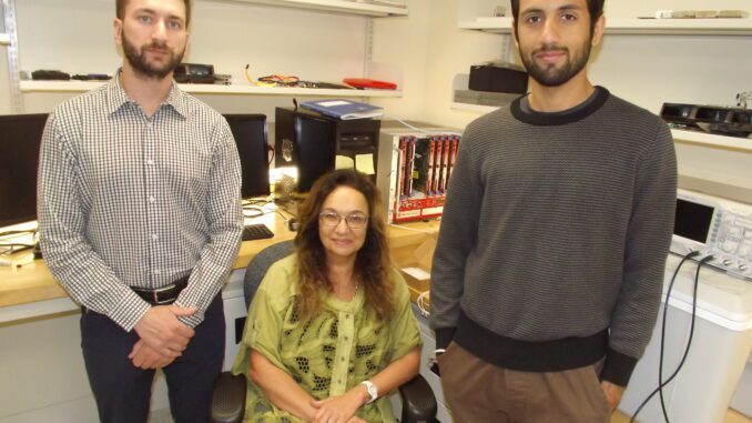
Photo – Group shot of 3 researchers:
Dr. Alla Reznik, centre, has been working on a medical imaging device that can be used to detect cancer in different parts of the body. She is pictured with Brandon Baldassi, left, and master’s student Anirudh Shahi.
Julio Heleno Gomes photo

Lakehead University is a Member of Research Canada: An Alliance for Health Discovery and a Sponsor of the Parliamentary Health Research Caucus Luncheon Celebrating the 2023 Gairdner Awards and Aligning First Nations’ Values in Indigenous Mental Health, Substance Use, Trauma and General Wellness: A Virtual Discussion with Dr. Christopher Mushquash. Visit rc-rc.ca to learn more.
